Box 4
Folder 25. Whales Fossil
Item 5. Ankle Morphology of the Earliest Cetaceans
![]()

Transcribed Text (OCR)
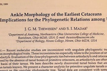
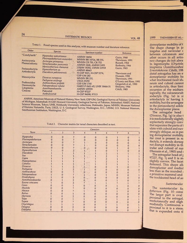
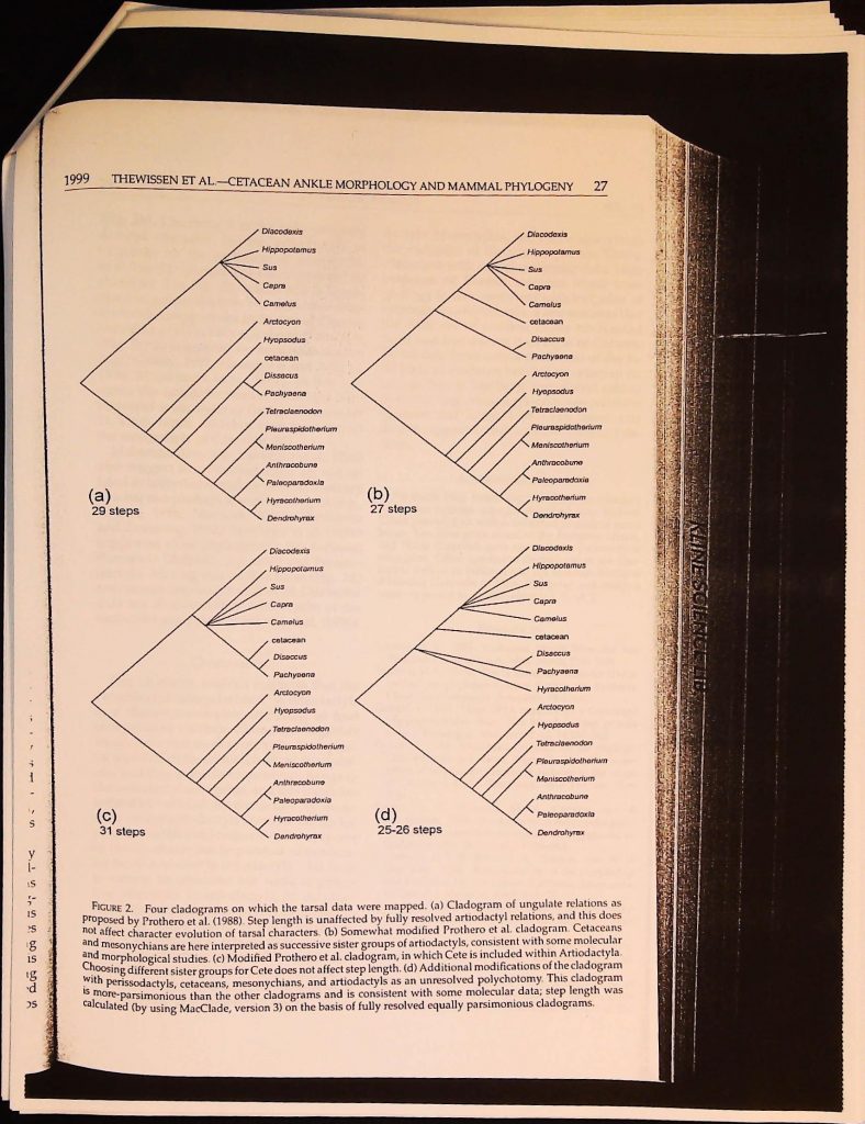
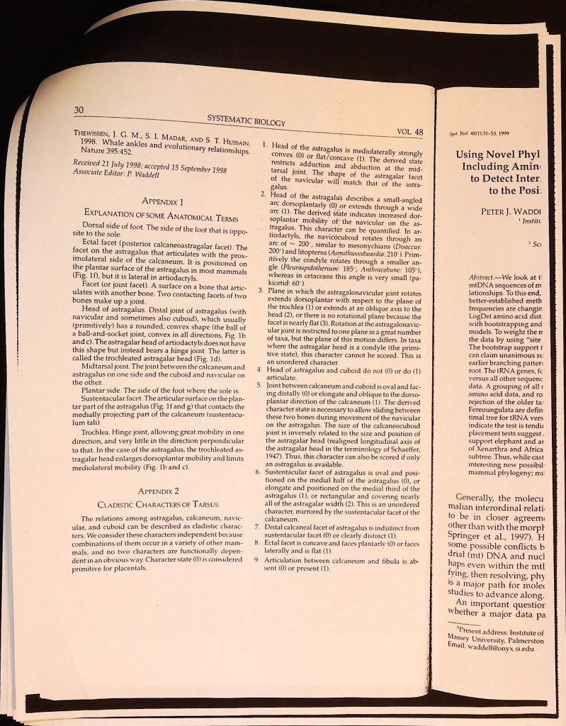
GARY MANGIACOPA ARCHIVE
============================================================
Title:      B4F25I5
Slug:       b4f25i5
Categories: Cryptozoology
Source:     https://garymangiacopraarchive.com/b4f25i5
Pages:      10 scanned, 10 extracted
OCR:        Google Vision API (document_text_detection)
Processed:  2026-06-06
============================================================

Syst. Biol. 48(1):21-30, 1999
Ankle Morphology of the Earliest Cetaceans
and Its Implications for the Phylogenetic Relations among Ungulates
J. G. M. THEWISSEN' AND S. I. MADAR²
'Department of Anatomy, Northeastern Ohio Universities College of Medicine,
Rootstown, Ohio 44240, USA; E-mail: thewisse@neoucom.edu
Department of Biology, Hiram College, Hiram, Ohio 44234, USA
Abstract. Recent molecular studies are inconsistent with ungulate phylogenetic trees that are
based on morphological traits. These inconsistencies especially relate to the position of cetaceans and
perissodactyls. Evaluation of the close phylogenetic ties between artiodactyls and cetaceans has been
hampered by the absence of tarsal bones of primitive cetaceans, as artiodactyls are often diagnosed
on the basis of their tarsus. We here describe newly discovered tarsal bones that are the oldest
cetacean tarsals known. We present a character analysis for primitive ungulate tarsals and evaluate
their impact on the ungulate phylogenetic tree. Tarsal data are consistent with some molecular
studies in suggesting that the extant sister group of Cetacea is Artiodactyla or that Cetacea should
be included within the latter order. Tarsal data do not support Cete (Mesonychia plus Cetacea)
and are consistent with the exclusion of perissodactyls from paenungulates as suggested by some
molecular studies. [Artiodactyla; Cetacea; locomotion; Mesonychia; Ungulata.]
The ankle (tarsus) plays a crucial role
in the morphological characterization of a
number of higher-level clades of mammals.
One of the main characters defining Pro-
boscidea, for instance, is the presence of
a medial process on the astragalus (Tassy,
1996). Archonta (Primates, Dermoptera,
Chiroptera, and Scandentia) are charac-
terized by fused sustentacular and nav-
icular facets on their astragalus (Szalay
and Drawhorn, 1980; for an explanation of
anatomical terms, see Appendix 1) and all
Artiodactyla bear a trochleated astragalar
head (Schaeffer, 1947). Despite major mor-
phological differences in ankle morphology
at higher taxonomic levels (e.g., Matthew,
1937), only minor differences commonly
occur within mammalian orders (e.g., ar-
tiodactyls: Hussain et al., 1983; Martinez
and Sudre, 1995). This makes the complex
of characters relating to ankle morphology
useful for phylogenetic analysis of higher
taxa.
One of the problems with phylogenetic
analyses based on tarsal morphology is
the delineation of cladistic characters from
a morphological continuum. Phylogenetic
characters should be consistent and inde-
pendent, and the delineation of morpho-
logical characters greatly affects cladogram
topology. We believe that functional studies
greatly elucidate the way in which char-
can
acters are delineated and do so in this study.
Central to our study is the shape of the
artiodactyl astragalus. The astragalar mor-
phology of artiodactyls is unique and plays
an important role in locomotor function
(Schaeffer, 1947, 1948). However, its unusual
function is the result of several morpholog-
ical features, a variable subset of which oc-
casionally occurs in other mammals as well.
Therefore, astragalar morphology cannot be
considered a single character, a point recog-
nized by Schaeffer (1948) but not by most
subsequent authors.
21
Because of its importance in character-
izing artiodactyls, astragalar morphology
plays a crucial role in determining the phy-
logenetic affinities of cetaceans. Cetaceans
have been placed within artiodactyls on the
basis of a large amount of recent molecular
data (Gatesy et al., 1996; Gatesy, 1997, 1998;
Hasegawa and Adachi, 1996; Shimamura
et al., 1997; Milinkovitch et al., 1998). This
view is commonly disputed by morpholog-
ical studies (Prothero et al., 1988; Thewis-
sen, 1994; Geisler and Luo, 1998; Luckett
and Hong, 1998; O'Leary, 1998; see also
Milinkovitch and Thewissen, 1997). Here,
we describe fragmentary astragali for pa-
kicetid and ambulocetid cetaceans (for dis-
cussion of these families, see Thewissen et
al., 1996, and Thewissen and Hussain, 1998).
These bones were previously unknown in

[PAGE BREAK]

22
SYSTEMATIC BIOLOGY
primitive cetaceans and have only been
mentioned in a short report (Thewissen et
al., 1998). These bones fill a major gap in
character matrices used to study the higher
phylogeny of ungulates, making it possible
to score cetaceans for several previously un-
known characters in the tarsus.
We here present a character analysis of the
complex of joints between the mammalian
astragalus, calcaneum, cuboid, and navic-
ular (Fig. 1). The midtarsal joint (anatomi-
cal terms are explained in Appendix 1) is of
specific interest. This complex joint involves
four bones; the trochleated head of the ar-
tiodactyl astragalus is part of the midtarsal
joint. Mobility at the midtarsal joint, how-
ever, is determined not only by the shape of
the joint facets that make up this joint but
also their relation to the ectal, sustentacu-
lar, and distal calcaneocuboid joints. These
facets sometimes lock the bones that partic-
ipate in the midtarsal joint at the point of
contact between calcaneum and astragalus.
Our description focuses on a primitive
ungulate (Pleuraspidotherium), artiodactyls,
and early whales, but we also consider the
morphology of a broader range of placen-
tals. There is relatively good consensus on
the morphology of the primitive placental
tarsus. Although different taxa were used to
identify the placental morphotype by dif-
ferent authors (Choeroclaenus by Schaeffer,
1947; Protungulatum by Szalay and Decker,
1974; Arctocyon by Cifelli, 1983), all are rel-
atively similar in the characters as delin-
eated here. The great age of these taxa and
their retention of primitive traits are also
consistent with their basal position on most
cladograms. We use the astragalus of the ar-
chaic ungulate Pleuraspidotherium (Thewis-
sen, 1991) as a basis for our description and
assume that most of its character scores are
plesiomorphic with respect to modern un-
gulates. This assumption is not critical for
our results, however, because we do not con-
struct a cladogram based on our limited data
set; we only map our characters on existing
cladograms.
We scored characters for the fossil taxa
listed in Table 1 as well as for several ex-
tant forms. Scores are reported in Table 2,
and the taxa used represent a cross-section
VOL. 48
1999
THEWISSEN ET AI
of all mammals as a reference for delineat-
ing characters and to facilitate comparisons.
We map these characters on a cladogram
that has been proposed for ungulates, recog-
nizing that, although these characters con-
tribute to the phylogenetic resolution within
ungulates, a complete phylogenetic analy-
sis should rely on all available data, not just
those of the ankle. Finally, we assess the
tarsal data in light of the conflict between
morphological and molecular studies of the
ungulate phylogenetic tree.
DESCRIPTION
Midtarsal Joint
The astragalar head of Pleuraspidotherium
(Fig. 1b) is strongly convex mediolater-
ally and dorsoplantarly. This permits some
movement between astragalus and navicu-
lar in all directions with no obvious axis of
mobility. Movement at this joint is limited in
part by the remainder of the midtarsal joint.
The plane of the joint between calcaneum
and cuboid in Pleuraspidotherium and other
primitive mammals is more proximal than
that between astragalus and navicular (Sza-
lay and Decker, 1974), which is not the case
in other placentals (Fig. 1a). The cuboid facet
of the calcaneum is rounded in outline prim-
itively, is mediolaterally and dorsoventrally
concave, and faces distally. This mismatch in
shapes between the astragalonavicular ar-
ticulation on the one hand and the calca-
neocuboid articulation on the other (Fig. 1b)
limits the degree of mobility of the midtarsal
joint. The cuboid does not articulate with the
astragalus.
The head of the astragalus in artiodactyls
(Fig. 1d) has the shape of a broad trochlea
(Schaeffer, 1947). This shape increases dor-
soplantar mobility of the navicular on the
astragalus. In addition, mediolateral mobil-
ity is completely checked by a parasagittal
groove that locks the navicular onto the as-
tragalus.
The calcaneocuboid joint of artiodactyls
is also modified (Fig. 1d). It is narrow, re-
stricted laterally, and slopes strongly dor-
sodistally. This shape allows the cuboid to
slide on the calcaneum while the navicu-
lar moves on the astragalus and increases
ASTRAGALUS
CA
CUNET
FORMES
NA
(a)
(f)
(i)
FIGURE 1.
NV V
-CU
ECTAI
SUST.
(c
SL
DIST.AS
Left tarsal bon
astragalus and calcaneum of,
tiodactyla), and Hyracotheriu
(Mesonychia), Hyracotherium
m) Dorsal view of astragalus
Table 1. Drawings are not to
numbers. Calc-cub= calcaneo
facet.

[PAGE BREAK]

1999
THEWISSEN ET AL. CETACEAN ANKLE MORPHOLOGY AND MAMMAL PHYLOGENY 23
ASTRAGALUS
CALCANEUM
CUNET
FORMES
(a)
II III
N V
(b)
NAVICULAR
CUBOID
TROCHLEA
HEAD
HEAD
(c)
ECTAL
(d)
CALC.-CUB.-
(e)
SUST.
et
1-
(f)
(g)
SUST.
n
r-
a-
b)
.al
(k)
(h)
he
ls
ea
or-
he
›il-
tal
as-
yls
re-
lor-
1 to
(CU-
Ises
(i)
FIGURE 1.
DIST.ASTR.
(j)
(1)
HEAD
(m)
Left tarsal bones of placental mammals. (a) Tarsus of a dog in dorsal view. (b-e) Distal view of
astragalus and calcaneum of, respectively, Pleuraspidotherium (condylarth), Oryctolagus (Lagomorpha), Capra (Ar-
tiodactyla), and Hyracotherium (Perissodactyla). (f-j) Plantar view of astragalus of Pleuraspidotherium, Dissacus
(Mesonychia), Hyracotherium, Diacodexis (Artiodactyla), and pakicetid cetacean (H-GSP 97227), respectively. (k-
m) Dorsal view of astragalus of Dissacus, pakicetid, and Diacodexis, respectively. Specimen numbers are listed in
Table 1. Drawings are not to scale and some are reversed from right elements. Roman numerals refer to digit
numbers. Calc-cub= calcaneocuboid facet; dist astr= distal astragalar facet; ectal=ectal facet; sust= sustentacular
facet.

[PAGE BREAK]

24
SYSTEMATIC BIOLOGY
TABLE 1. Fossil species used in this analysis, with museum number and literature reference.
Order
"Condylarth"
Arctocyonia
Phenacodonta
Perissodactyla
Artiodactyla
Mesonychia
Proboscidea
Desmostylia
Litopterna
Cetacea
Species
Hyopsodus walcottianus
Pleuraspidotherium aumonieri
Arctocyon primaevus
Tetraclaenodon puercensis
Meniscotherium chamense
Hyracotherium sp.
Diacodexis pakistanensis
Dissacus europaeus
Pachyaena ossifraga
Anthracobune pinfoldi
Paleoparadoxia tubatai
Asmithwoodwardia
Pakicetid
Ambulocetid
Specimen number
AMNH 14654
MNHN BR 10726, BR 55L
MNHN CR 750, CR 752
AMNH 2468, AMNH 2493
USNM 18282, USNM 22918
AMNH 48663
H-GSP 5021, H-GSP 5274,
GSP-UM 282
MNHN BR 21L
USGS 25292
GSP-UM 1745
NMST-P-5601-45, UHR 18466-31
AMNH 109555
H-GSP 97227
H-GSP 96098, 97113
VOL. 48
Reference
Gazin, 1968
Thewissen, 1991
Russell, 1964
Radinsky, 1966
Gazin, 1965
Thewissen and
Hussain, 1990
Thewissen, 1991
O'Leary and Rose, 1995
Gingerich et al., 1990
Shikama, 1966
Cifelli, 1983
AMNH, American Museum of Natural History, New York; GSP-UM, Geological Survey of Pakistan-University
of Michigan, Islamabad; H-GSP, Howard University, Geological Survey of Pakistan, Islamabad; NMST, National
Science Museum, Tokyo; UHR, Hokkaido University collections, Hokkaido, Japan; MNHN, Museum National
d'Histoire Naturelle, Paris, USGS, U. S. Geological Survey, Washington, D.C., USNM, U.S. National Museum,
Smithsonian Institution, Washington, D.C.
TABLE 2.
Character matrix for tarsal characters described in text.
oooooo
Characters
Taxon
Hyopsodus
Pleuraspidotherium
Arctocyon
Tetraclaenodon
Meniscotherium
Hyracotherium
Diacodexis
Sus
Capra
Hippopotamus
Camelus
Dissacus
Pachyaena
Anthracobune
Paleoparadoxia
Dendrohyrax
Asmithwoodwardia
Eocene cetaceans
Canis
Mustela
Macaca
Rattus
Marmota
Talpa
Tupaia
Oryctolagus
Dasypus
Didelphis
boooo0000
000100010
12222323122121212.
100111001001¯¯¯o
ooooooooo
1999
THEWISSEN ET AL.-
dorsoplantar mobility of t
The shape change in jo
tragalus and navicular c
between calcaneum and
other occurs in all artio
two changes do not alwa
In lagomorphs (Oryctola
litopterns (Asmithwoodwa
Ernestokokenia: Cifelli, 198
distal astragalus has an e
dorsoplantar mobility ba
what trochleated facet sha
neum and cuboid cannot
in artiodactyls, thus limi
excursion at the midtars
logically, the calcaneocub
sodactyls (Fig. 1e) is si
artiodactyls in having la
mobility, but the arrangem
in the perissodactyl ankle
the dorsoplantar plane.
The astragalar head
(Dissacus, Fig. 1g) is also t
it is mediolaterally slightly
soplantarly strongly conv
that separates the parts of t
ulate with cuboid and nav
strongly oblique, as in per
ing dorsoplantar mobility
ilar crest is present in no
dactyls, it extends dorsop
not disrupt mobility in th
icular and cuboid of run
(Hussain et al., 1983) and
The astragalar head of
97227, Fig. 1j and 1) is m
slightly convex. The facet
flattened. This shape all
dorsoplantar and medio!
less than in the rounded :
a primitive mammal and
the trochleated head of an
Sustentacular
The sustentacular fa
dotherium (Fig. 1f) consi
The larger part is oval
proximal to the astragal.
mediolaterally and sligh
modistally. Continuous w
proximal to it is a stron
that is expanded onto th

[PAGE BREAK]

.5
ty
al
al
n,
1910
1999
THEWISSEN ET AL. CETACEAN ANKLE MORPHOLOGY AND MAMMAL PHYLOGENY 25
dorsoplantar mobility of the midtarsal joint.
The shape change in joints between as-
tragalus and navicular on one hand and
between calcaneum and cuboid on the
other occurs in all artiodactyls but these
two changes do not always occur together.
In lagomorphs (Oryctolagus, Fig. 1c) and
litopterns (Asmithwoodwardia, synonym of
Ernestokokenia: Cifelli, 1983), the head of the
distal astragalus has an expanded range of
dorsoplantar mobility based on its some-
what trochleated facet shape, but the calca-
neum and cuboid cannot slide as they do
in artiodactyls, thus limiting dorsoplantar
excursion at the midtarsal joint. Morpho-
logically, the calcaneocuboid joint of peris-
sodactyls (Fig. 1e) is similar to that of
artiodactyls in having laterally restricted
mobility, but the arrangement of other facets
in the perissodactyl ankle limits mobility in
the dorsoplantar plane.
The astragalar head of mesonychians
(Dissacus, Fig. 1g) is also trochleated in that
it is mediolaterally slightly concave and dor-
soplantarly strongly convex. It bears a crest
that separates the parts of the head that artic-
ulate with cuboid and navicular. This crest is
strongly oblique, as in perissodactyls, limit-
ing dorsoplantar mobility. Although a sim-
ilar crest is present in nonruminant artio-
dactyls, it extends dorsoplantarly and does
not disrupt mobility in this plane. The nav-
icular and cuboid of ruminants are fused
(Hussain et al., 1983) and no crest is present.
The astragalar head of pakicetids (H-GSP
97227, Fig. 1j and 1) is mediolaterally only
slightly convex. The facet is dorsoplantarly
flattened. This shape allows very limited
dorsoplantar and mediolateral mobility-
less than in the rounded astragalar head of
a primitive mammal and much less than in
the trochleated head of an artiodactyl.
Sustentacular Facet
The sustentacular facet of Pleuraspi-
dotherium (Fig. 1f) consists of two parts.
The larger part is oval and located just
proximal to the astragalar head; it is flat
mediolaterally and slightly convex proxi-
modistally. Continuous with this part and
proximal to it is a strongly concave facet
that is expanded onto the medial half of
the plantar surface of the trochlea. The con-
cavity of this part is oblique to the long
axis of the bone and has the same axis as
the concavity of the ectal facet. Combined,
these two facets limit the mobility of the
calcaneum on the astragalus to a direction
oblique to the long axis of the foot. The
sustentacular facet of Pleuraspidotherium is
not continuous with the articular surface of
the astragalar head. The sustentacular facet
of plantigrade mammals, such as Pleuraspi-
dotherium, has a weight-bearing function,
transmitting force from the astragalus to the
calcaneum.
The sustentacular facet of artiodactyls
(Fig. 1i) is greatly expanded. Its mediolateral
profile may be concave or convex, but it is
always convex proximodistally and the
main axis of mobility is dorsoplantar. The
sustentacular facet of artiodactyls covers
most of the width of the plantar side of the
astragalus.
In mesonychians, the sustentacular facet
is similar to that of Pleuraspidotherium, in
that it consists of a concave and a convex
part that limit mobility between astragalus
and calcaneum. The sustentacular facet of
pakicetid cetaceans (H-GSP 97227) is long
and narrow, limited to the medial third of the
astragalus, but it is proximodistally convex,
as in artiodactyls. Only a small fragment
of the sustentacular facet of the ambulo-
cetid astragalus (H-GSP 96098) is preserved.
Its morphology is consistent with that of
the pakicetid.
Ectal Facet
The ectal facet of Pleuraspidotherium
(Fig. 1f) faces plantarly and is elongate
and strongly concave along an oblique axis.
This concavity matches that of the sus-
tentacular facet and locks the ectal facet
of the astragalus onto the calcaneum, lim-
iting mobility at the midtarsal joint. In
perissodactyls (Fig. 1h), litopterns, and
lagomorphs, the axis of the ectal facet ex-
tends more mediolaterally than in prim-
itive astragali (Fig. 1f), limiting dorso-
plantar mobility between astragalus and
calcaneum.
The ectal facet of artiodactyls is highly
modified. It is narrow and lies on the lateral

[PAGE BREAK]

26
SYSTEMATIC BIOLOGY
side of the calcaneum (and is hence not vis-
is strongly reduced and variable in outline.
ible in Fig. li). The ectal facet of artiodactyls
This reduction permits astragalar rotation
independent of the calcaneum (Schaeffer,
1947).
The ectal facet of mesonychians (Fig. 1g)
is similar to that of Pleuraspidotherium. The
pakicetid astragalus does not preserve the
ectal facet, but the proximal part of the am-
bulocetid astragalus (H-GSP 96098) is well-
preserved. Its ectal facet is laterally placed
(see Thewissen et al., 1998). No part of the
and resembles artiodactyls in morphology
ectal facet extends on the plantar surface of
the astragalus.
FUNCTIONAL INFERENCE
The artiodactyl midtarsal joint is uniquely
mobile dorsoplantarly but is immobile
mediolaterally as a result of extensive re-
modeling of the ankle. This remodeling ap-
pears to consist of four independent cladis-
tic characters, none of which is unique to
artiodactyls. First, the astragalar head is
expanded dorsoplantarly (character 2, Ap-
pendix 2), as is also the case in litopterns
and mesonychians. Second, the plane of
rotation of the astragalonavicular joint is
dorsoplantar (character 3), as occurs also
in lagomorphs and dasypodids. Third, the
calcaneocuboid joint is narrow and ex-
tends from proximoplantar to distodorsal
on the lateral side of the foot (charac-
ter 5), similar to perissodactyls. Fourth,
the ectal facet is small and faces later-
ally (character 8), as it does in primitive
cetaceans. Artiodactyls share all four char-
acters, but litopterns, mesonychians, lago-
morphs, dasypodids, perissodactyls, and
cetaceans have only some of the characters
and their midtarsal joints are not highly mo-
bile.
Increased dorsoplantar mobility in artio-
dactyls is combined with enhanced stability
mediolaterally. This is related to two fea-
tures of the heel. First, movement at the
astragalar head is restricted mediolater-
ally (character 1), as it is in perissodactyls,
mesonychians, desmostylians, hyracoids,
lagomorphs, and primitive cetaceans. Sec-
ond, the sustentacular facet is expanded to
VOL. 48
1999
THEWISSEN ET AL.-CET
cover much of the plantar face of the astra-
galus (character 6). This character is unique
to artiodactyls.
PHYLOGENETIC INFERENCE
The modern orders of ungulates have
a long independent history, and rever-
sals and autapomorphies that accumulated
since their divergence may seriously af-
fect character analyses. The absence of the
tarsus in modern cetaceans is an excel-
lent example of such an autapomorphy.
To avoid problems of homoplasy during
their independent evolution, we studied
as many Paleogene representatives of or-
ders as possible. Because our set of char-
acters is limited to those in the tarsus (Ta-
ble 2), we did not execute a phylogenetic
analysis, but instead mapped our charac-
ters on a published phylogeny. We chose
the phylogeny of Prothero et al. (1988) be-
cause it includes nearly all of the rele-
vant taxa and is fully resolved. Overall,
this analysis is relatively similar to more
broad-scale morphological analyses based
on modern taxa (e.g., Gaudin et al., 1996).
The Prothero et al. phylogeny (Fig. 2a)
was not based on an explicit phyloge-
netic analysis and thus may not be the
most-parsimonious topology, but it is use-
ful as a starting point for our study. Map-
ping our tarsal data on this phylogeny
indicates that 29 character state changes
took place. Most notable among the char-
acter changes implied in this phylogeny
is character 8. The derived state of this
character is the only feature that could
be interpreted as a unique synapomor-
phy of cetaceans and artiodactyls; instead,
it is treated as a homoplasious in this
analysis.
Recent molecular data sets (reviewed by
Gatesy, 1998) are incongruent with the topol-
ogy of Figure 2a and suggest that cetaceans
are allied with artiodactyls. Amending Fig-
ure 2a to make Cete (Mesonychia plus
Cetacea) the sister group of artiodactyls does
not decrease the step length, but moving
only Cetacea to the artiodactyl branch is
more parsimonious (27 steps). Considering
Mesonychia as the sister group to combined
Cetacea plus Artiodactyla also costs 27 steps
(a)
29 steps
(c)
31 steps
FIGURE 2.
Four cladograms on w
proposed by Prothero et al. (1988). St
not affect character evolution of tars
and mesonychians are here interprete
and morphological studies. (c) Modit
Choosing different sister groups for C
with perissodactyls, cetaceans, meso
is more-parsimonious than the othe
calculated (by using MacClade, versi

[PAGE BREAK]

1999
THEWISSEN ET AL-CETACEAN ANKLE MORPHOLOGY AND MAMMAL PHYLOGENY
S
VESTSS
15
g
IS
OS
Dlacodexis
Hippopotamus
Sus
Capra
Camelus
Arctocyon
Hyopsodus
cetacean
Diacodexis
Hippopotamus
Sus
Capra
Camelus
cotacoan
Disaccus
Pachysona
Arctocyon
27
(a)
29 steps
Dissacus
Pachyaena
Tetraclaenodon
Plauraspidotherium
Meniscotherium
Anthracobune
Paleoparadoxia
Hyracotherium
Dendrohyrax
(b)
27 steps
Hyopsodus
Tetraclaenodon
Pleuraspidotherium
Meniscotherium
Anthracobuna
Paleoparadoxia
Hyracotherium
Dendrohyrax
(c)
31 steps
Diacodexis
Hippopotamus
Sus
Capra
Camelus
cetacean
Disaccus
Pachyaena
Arclocyon
Hyopsodus
Tetraclaenodon
Pleuraspidotherium
Meniscotherium
Anthracobune
Paleoparadoxia
Hyracotherium
Dandrohyrax
Diacodexis
Hippopotamus
Sus
Capra
Camelus
cetacean
Disaccus
Pachyaena
Hyracotherium
Arctocyon
Hyopsodus
Tetraclaenodon
Plouraspidothorium
Moniscolherium
Anthracobune
(d)
25-26 steps
Paleoparadoxla
Dendrohyrax
FIGURE 2. Four cladograms on which the tarsal data were mapped. (a) Cladogram of ungulate relations as
proposed by Prothero et al. (1988). Step length is unaffected by fully resolved artiodactyl relations, and this does
not affect character evolution of tarsal characters. (b) Somewhat modified Prothero et al. cladogram. Cetaceans
and mesonychians are here interpreted as successive sister groups of artiodactyls, consistent with some molecular
and morphological studies. (c) Modified Prothero et al. cladogram, in which Cete is included within Artiodactyla.
Choosing different sister groups for Cete does not affect step length. (d) Additional modifications of the cladogram
with perissodactyls, cetaceans, mesonychians, and artiodactyls as an unresolved polychotomy. This cladogram
is more-parsimonious than the other cladograms and is consistent with some molecular data; step length was
calculated (by using MacClade, version 3) on the basis of fully resolved equally parsimonious cladograms.

[PAGE BREAK]

28
SYSTEMATIC BIOLOGY
derived character linking artiodactyls to
(Fig. 2b). Character 8 is the only uniquely
cetaceans (to the exclusion of mesonychi-
character 5 in Fig. 2b, where it is a homo-
ans), but this grouping is also supported by
plasy in perissodactyls. The same two char-
acters also support the inclusion of Cetacea
in Artiodactyla at a branch above Diacodexis,
(e.g., Gatesy, 1997; Hasegawa and Adachi,
(Fig. 2c) as suggested by molecular studies
1996; Shimamura et al., 1997). In this sce-
nario, the astragalar head of cetaceans is in-
terpreted as secondarily flattened (character
2). Thus, tarsal evidence does not support
the monophyly of Cete (Fig. 2c).
Molecular data (e.g., Lavergne et al., 1996)
also suggest that Perissodactyla are not re-
lated to tethytheres and hyracoids, contrary
to what morphological studies suggest (e.g.,
Fischer, 1986; Novacek, 1992; Thewissen
and Domning, 1992). Stanhope et al. (1996)
supported close phylogenetic ties between
Paraxonia (dolphin, pig, cow) and Perisso-
dactyla. Making this modification (Fig. 2d)
reduces the step length to 25 or 26. Our tarsal
data are thus consistent with results of the
molecular analysis of Stanhope et al. (1996).
CONCLUSIONS
Our character analysis suggests that the
is a character complex consisting of four
"trochleated astragalar head" of artiodactyls
independent cladistic characters. Together
these characters work to increase dorsoplan-
tar mobility of the tarsus. Limiting mediolat-
eral mobility is another functional change in
the artiodactyl astragalus, and this is charac-
terized by two cladistic characters. No mam-
mals
except artiodactyls combine the six (or
even the initial four) characters, but only one
of the six characters (expanded sustentacu-
lar facet) is unique to artiodactyls. This facet
is narrow in cetaceans but wide in primi-
tive placentals. In artiodactyls, on the other
hand, the sustentacular facet is expanded to
cover the entire width of the plantar sur-
face. Cetaceans and artiodactyls share the
derived position and shape of the ectal facet,
a condition not shared by mesonychians.
We conclude that tarsal data support close
phylogenetic ties between Cetacea, Artio-
VOL. 48
dactyla, Mesonychia, and Perissodactyla, to
the exclusion of the paenungulates. This is
consistent with recent molecular data. New
evidence of Eocene cetacean tarsal mor-
phology is also consistent with inclusion of
cetaceans in artiodactyls, if one assumes that
the wide arc of rotation of the trochleated
head was lost during the origin of Cetacea.
Tarsal data form only a small part of the to-
tal body of evidence bearing on cetacean re-
lations, but it has been considered critical
in evaluating artiodactyl-cetacean relations
(Luckett and Hong, 1998). The tarsal data
do not support mesonychian-cetacean rela-
tions.
ACKNOWLEDGMENTS
We thank Tony Friscia and Lois Roe, the collectors
of the two cetacean astragali described here. Richard
Cifelli provided us with a collection of casts of tarsal
bones, Ken Rose gave access to the Pachyaena tarsals,
and Philip Gingerich gave access to the Anthracobune
astragalus. Other specimens studied are at the Geo-
logical Survey of Pakistan and the Museum National
d'Histoire Naturelle in Paris. Fieldwork and analysis
were supported by an NSF grant to J. G. M. T.
REFERENCES
CIFELLI, R. L. 1983. Eutherian tarsals from the late
Paleocene of Brazil. Am. Mus. Novit. 2761:1-31.
FISCHER, M. S. 1986 Die Stellung der Schliefer (Hyra-
coidea) im phylogenetischen System der Eutheria.
Cour. Forschungsinst. Senckenb. 84:1-132.
GATESY, J. 1997. More DNA support for a Cetacean/
Hippopotamidae clade: The blood-clotting protein
gene y-fibrinogen. Mol. Biol. Evol. 14:537-543.
GATESY, J. 1998. Molecular evidence for the phyloge
netic affinities of Cetacea Pages 63-111 in The emer-
gence of whales: Evolutionary patterns in the origin
of Cetacea (J. G. M. Thewissen, ed.). Plenum, New
York.
GATESY, J., C. HAYASHI, M. A. CRONIN, AND P.
ARCTANDER. 1996. Evidence from milk casein genes
that cetaceans are close relatives of hippopotamid ar-
tiodactyls. Mol. Biol. Evol. 13:954-963.
GAUDIN, T. J., J. R. WIBLE, J. A. HOPSON, AND W. D.
TURNBULL. 1996. Reexamination of the morpholog-
ical evidence for the cohort Epitheria (Mammalia,
Eutheria). J. Mammal Evol. 3:31-77.
GAZIN, C. L. 1965. A study of the early tertiary condy-
larthran mammal Meniscotherium. Smithson. Misc.
Collect. 149:1-98.
GAZIN, C. L. 1968. A study of the Eocene condy-
larthran mammal Hyopsodus. Smithson. Misc. Col-
lect. 153:1-90.
GEISLER, J. H., AND Z. Luo. 1998. Relationships of
Cetacea to terrestrial ungulates and the evolution
of cranial vasculature in Cete. Pages 163-212 in The
1999
THEWISSEN ET AL.-
emergence of whales: Evolutio
origin of Cetacea (J. G. M. Thev
New York.
GINGERICH, P. D., D. E. RUSSELL,
1990. Astragalus of Anthrac
Proboscidea) from the early.
Kashmir. Contrib. Mus. Pale
28:71-77.
HASEGAWA, M., AND J. ADACHI. 19
sition of cetaceans relative to art:
of mitochondrial and nuclear s
Evol. 13:710-717.
HUSSAIN, S. T., P. Y. SONDAAR,
G. M. THEWISSEN, E. F. H. M.
SPOOR. 1983. Fossil mammal
field atlas. Part I: The artiodact
Geol. Surv. Pak. 14:1-14.
LAVERGNE, A., E. DOUZERY, T. STICH
AND M. S. SPRINGER. 1996.
malian relationships: Evidenc
monophyly is provided by c
drial 125 rRNA sequences. Mo
6:245-258.
LUCKETT, W. P., AND N. HONG. 19°
lationships between the order
Cetacea: A combined assessm
ical and molecular evidence.
5:127-182.
MARTINEZ, J.-N., AND J. SUDRE. 1
of Paleogene artiodactyls: Co
ogy, variability and prediction o
28.197-209.
MATTHEW, W. D. 1937. Paleocen
Juan Basin. Trans. Am. Philos. S
MILINKOVITCH, M. C., M. BÉRUBÉ,
1998. Are cetaceans highly d
Pages 113-131 in The eme
Evolutionary patterns in the
(J. G. M. Thewissen, ed.). Plenu:
MILINKOVITCH, M. C., AND J. G. M
Even-toed fingerprints on wha
388:622-624.
NOVACEK, M. J. 1992. Fossils, top
and the higher level phylogeny
mals. Syst. Biol. 41:58-73.
O'LEARY, M. A. 1998. Phylogenet:
reassessment of the dental evic
chian and cetacean clade. Pages
gence of whales: Evolutionary
of Cetacea (J. G. M. Thewissen
York.
O'LEARY, M. A., AND K. D. Ros
skeleton of the early Eocene m
(Mammalia, Mesonychia). J. V
430.
PROTHERO, D., E. MANNING, AND M
phylogeny of ungulates. Pages
logeny and classification of the
Mammals (M. Benton, ed.). Clar
England.
RADINSKY, L. B. 1966. The adapt
phenacodontid condylarths an
Perissodactyla. Evolution 20:40

[PAGE BREAK]

KLINE SCIENCE LIB
1999
THEWISSEN ET AL. CETACEAN ANKLE MORPHOLOGY AND MAMMAL PHYLOGENY
emergence of whales: Evolutionary patterns in the
origin of Cetacea (J. G. M. Thewissen, ed.). Plenum,
New York
GINGERICH, P. D., D. E. RUSSELL, AND N. A. WELLS.
1990. Astragalus of Anthracobune (Mammalia,
Proboscidea) from the early-middle Eocene of
Kashmir. Contrib. Mus. Paleontol. Univ. Mich.
28 71-77.
HASEGAWA, M., AND J. ADACHI. 1996. Phylogenetic po-
sition of cetaceans relative to artiodactyls: Reanalysis
of mitochondrial and nuclear sequences. Mol. Biol.
Evol. 13:710-717.
HUSSAIN, S. T., P. Y. SONDAAR, S. M. I. SHAH, J.
G. M. THEWISSEN, E. F. H. M. COUSIN, AND C. F.
SPOOR. 1983. Fossil mammal bones of Pakistan, a
field atlas. Part I: The artiodactyl astragalus. Mem.
Geol Surv. Pak. 14:1-14.
LAVERGNE, A., E. DOUZERY, T. STICHLER, F. M. CATZEFLIS,
AND M. S. SPRINGER. 1996. Interordinal mam-
malian relationships: Evidence for paenungulate
monophyly is provided by complete mitochon-
drial 125 rRNA sequences. Mol. Phylogenet. Evol
6:245-258.
LUCKETT, W. P., AND N. HONG. 1998. Phylogenetic re-
lationships between the orders Artiodactyla and
Cetacea: A combined assessment of morpholog-
ical and molecular evidence. J. Mammal Evol.
5:127-182.
MARTINEZ, J.-N., AND J. SUDRE. 1995. The astragalus
of Paleogene artiodactyls: Comparative morphol-
ogy, variability and prediction of body mass Lethaia
28:197-209.
MATTHEW, W. D. 1937. Paleocene faunas of the San
Juan Basin. Trans. Am. Philos. Soc., N. S. 30:1-510.
MILINKOVITCH, M. C., M. BÉRUBÉ, AND P. J. PALSBØLL.
1998. Are cetaceans highly derived artiodactyls?
Pages 113-131 in The emergence of whales:
Evolutionary patterns in the origin of Cetacea
(J.G M. Thewissen, ed.). Plenum, New York.
MILINKOVITCH, M. C., AND J. G. M. THEWISSEN. 1997.
Even-toed fingerprints on whale ancestry. Nature
388.622-624.
NOVACEK, M. J. 1992. Fossils, topologies, missing data,
and the higher level phylogeny of eutherian mam-
mals. Syst. Biol. 41:58-73.
O'LEARY, M. A. 1998. Phylogenetic and morphometric
reassessment of the dental evidence for a mesony-
chian and cetacean clade Pages 133-162 in The emer-
gence of whales: Evolutionary patterns in the origin
of Cetacea (J. G. M. Thewissen, ed.). Plenum, New
York.
O'LEARY, M. A., AND K. D. ROSE. 1995. Postcranial
skeleton of the early Eocene mesonychid Pachyaena
(Mammalia, Mesonychia). J. Vert. Paleont. 15:401-
430.
PROTHERO, D., E. MANNING, AND M. FISCHER. 1988. The
phylogeny of ungulates. Pages 201-234 in The phy-
logeny and classification of the tetrapods, Volume 2.
Mammals (M. Benton, ed.). Clarendon Press, Oxford,
England.
RADINSKY, L. B. 1966. The adaptive radiation of the
phenacodontid condylarths and the origin of the
Perissodactyla. Evolution 20:408–417.
29
RUSSELL, D. E. 1964. Les mammiféres paléocènes
d'Europe. Mém. Mus. Natl. d'Hist. Nat. Ser. C 13:1-
324.
SCHAEFFER, B. 1947. Notes on the origin and func-
tion of the artiodactyl tarsus. Am. Mus. Novit.
1350:1-24.
SCHAEFFER, B. 1948. The origin of a mammalian ordi-
nal character. Evolution 2:164-175.
SHIKAMA, T. 1966. Postcranial skeletons of Japanese
Desmostylia, Palaeontol. Soc. Jpn. Spec. Pap. 12:1-
202.
SHIMAMURA, M, H. YASUE, K. OHSHIMA, H. ABE, H.
KATO, T. KISHRO, M. GOTO, I. MUNECHIKAL, AND
N. OKADA. 1997. Molecular evidence from retro-
posons that whales form a clade within even-toed
ungulates. Nature 388:666-670.
STANHOPE, M. J., M. R. SMITH, P. G. WADDELL, C. A.
PORTER, M. S. SHIVJI, AND M. GOODMAN. 1996. Mam-
malian evolution and the interphotoreceptor
retinoid binding protein (IRBP) gene: Convincing
evidence for several superordinal clades. J. Mol.
Evol. 43:83-92.
SZALAY, F. S., AND R. L. DECKER. 1974. Origins, evo-
lution of the tarsus in late Cretaceous eutheria and
Paleocene primates. Pages 223-259 in Primate Lo-
comotion (F. A. Jenkins, ed.). Academic Press, New
York.
SZALAY, F S., AND G. DRAWHORN. 1980. Evolution and
diversification of the Archonta in an arboreal milieu.
Pages 133-169 in Comparative biology and evolu-
tionary relationships of tree shrews (W. P. Luckett,
ed.). Plenum, New York.
TASSY, P. 1996. Who is who among Proboscidea?
Pages 39-54 in The Proboscidea: Evolution and
palaeoecology of elephants and their relatives
(J. Shoshani and P. Tassy, eds.). Oxford Univ. Press,
New York.
THEWISSEN, J. G. M. 1991. Limb osteology and func-
tion of the primitive Paleocene ungulate Pleuraspi-
dotherium with notes on Tricuspiodon and Dissacus
(Mammalia). Geobios 24:483–495.
THEWISSEN, J. G. M. 1994 (published in 1995). Phylo-
genetic aspects of cetacean origins: A morphological
perspective. J. Mammal. Evol. 2.157-184.
THEWISSEN, J. G. M., AND D. P. DOMNING. 1992. The role
of phenacodontids in the origin of the modern orders
of ungulate mammals. J. Vertebr. Paleontol. 12:494-
504.
THEWISSEN, J. G. M., AND S. T. HUSSAIN. 1990. Postcra-
nial osteology of the most primitive artiodactyl, Dia-
codexis pakistanensis (Dichobunidae). Anat. Hist. Em-
bryol. (Berl.) 19.37-48.
THEWISSEN, J. G. M., AND S. T. HUSSAIN. 1998. Sys-
tematic review of the Pakicetidae, early and mid-
dle Eocene Cetacea (Mammalia) from Pakistan and
India. Pages 220-238 in Dawn of the age of
mammals in Asia (C. Beard and M. R. Dawson, eds.).
Carnegie Museum of Natural History, Pittsburgh,
Pennsylvania.
THEWISSEN, J. G. M., S. I. MADAR, AND S. T. HUSSAIN.
1996. Ambulocetus natans, an Eocene cetacean
(Mammalia) from Pakistan. Cour. Forschungsinst.
Senckenb. 191:1-86.
of
n
e

[PAGE BREAK]

30
SYSTEMATIC BIOLOGY
THEWISSEN, J. G. M., S. I. MADAR, AND S. T. HUSSAIN.
Nature 395:452.
1998. Whale ankles and evolutionary relationships.
Received 21 July 1998; accepted 15 September 1998
Associate Editor: P. Waddell
APPENDIX 1
EXPLANATION OF SOME ANATOMICAL TERMS
site to the sole.
Dorsal side of foot. The side of the foot that is oppo-
Ectal facet (posterior calcaneoastragalar facet). The
facet on the astragalus that articulates with the prox-
imolateral side of the calcaneum. It is positioned on
the plantar surface of the astragalus in most mammals
(Fig. 1f), but it is lateral in artiodactyls.
Facet (or joint facet). A surface on a bone that artic-
ulates with another bone. Two contacting facets of two
bones make up a joint.
Head of astragalus. Distal joint of astragalus (with
navicular and sometimes also cuboid), which usually
(primitively) has a rounded, convex shape (the ball of
a ball-and-socket joint, convex in all directions, Fig. 1b
and c). The astragalar head of artiodactyls does not have
this shape but instead bears a hinge joint. The latter is
called the trochleated astragalar head (Fig. 1d).
Midtarsal joint. The joint between the calcaneum and
astragalus on one side and the cuboid and navicular on
the other.
Plantar side. The side of the foot where the sole is.
Sustentacular facet. The articular surface on the plan-
tar part of the astragalus (Fig. 1f and g) that contacts the
medially projecting part of the calcaneum (sustentacu-
lum tali).
Trochlea. Hinge joint, allowing great mobility in one
direction, and very little in the direction perpendicular
to that. In the case of the astragalus, the trochleated as-
tragalar head enlarges dorsoplantar mobility and limits
mediolateral mobility (Fig. 1b and c).
APPENDIX 2
CLADISTIC CHARACTERS OF TARSUS
The relations among astragalus, calcaneum, navic-
ular, and cuboid can be described as cladistic charac-
ters. We consider these characters independent because
combinations of them occur in a variety of other mam-
mals, and no two characters are functionally depen-
dent in an obvious way. Character state (0) is considered
primitive for placentals.
VOL. 48
1. Head of the astragalus is mediolaterally strongly
convex (0) or flat/concave (1). The derived state
restricts adduction and abduction at the mid-
tarsal joint. The shape of the astragalar facet
galus.
of the navicular will match that of the astra-
2. Head of the astragalus describes a small-angled
arc dorsoplantarly (0) or extends through a wide
arc (1). The derived state indicates increased dor-
soplantar mobility of the navicular on the as-
tragalus. This character can be quantified. In ar-
tiodactyls, the navicócuboid rotates through an
arc of ~200, similar to mesonychians (Disaccus:
200°) and litopterns (Asmithwoodwardia: 210). Prim-
itively the condyle rotates through a smaller an-
gle (Pleuraspidotherium 185°, Anthracobune: 105°),
whereas in cetaceans this angle is very small (pa-
kicetid: 60°)
3. Plane in which the astragalonavicular joint rotates
extends dorsoplantar with respect to the plane of
the trochlea (1) or extends at an oblique axis to the
head (2), or there is no rotational plane because the
facet is nearly flat (3). Rotation at the astragalonavic-
ular joint is restricted to one plane in a great number
of taxa, but the plane of this motion differs. In taxa
where the astragalar head is a condyle (the primi-
tive state), this character cannot be scored. This is
an unordered character.
4 Head of astragalus and cuboid do not (0) or do (1)
articulate.
5. Joint between calcaneum and cuboid is oval and fac-
ing distally (0) or elongate and oblique to the dorso-
plantar direction of the calcaneum (1). The derived
character state is necessary to allow sliding between
these two bones during movement of the navicular
on the astragalus. The size of the calcaneocuboid
joint is inversely related to the size and position of
the astragalar head (realigned longitudinal axis of
the astragalar head in the terminology of Schaeffer,
1947). Thus, this character can also be scored if only
an astragalus is available.
6. Sustentacular facet of astragalus is oval and posi-
tioned on the medial half of the astragalus (0), or
elongate and positioned on the medial third of the
astragalus (1), or rectangular and covering nearly
all of the astragalar width (2). This is an unordered
character, mirrored by the sustentacular facet of the
calcaneum.
7. Distal calcaneal facet of astragalus is indistinct from
sustentacular facet (0) or clearly distinct (1).
8. Ectal facet is concave and faces plantarly (0) or faces
laterally and is flat (1).
9. Articulation between calcaneum and fibula is ab-
sent (0) or present (1).
Syst. Biol 48(1).31-53, 1999
Using Novel Phyl
Including Amin
to Detect Inter.
to the Posi.
PETER J. WADDI
'Institu
: Sci
Abstract. We look at t
mtDNA sequences of m
lationships. To this end,
better-established meth
frequencies are changin
LogDet amino acid dist
with bootstrapping and
models. To weight the n
the data by using "site
The bootstrap support!
can claim unanimous su
earlier branching patter
root. The tRNA genes, fc
versus all other sequenc
data. A grouping of all
amino acid data, and ro
rejection of the older ta:
Fereuungulata are defin
timal tree for tRNA vers
indicate the test is tendi
placement tests suggest.
support elephant and ar
of Xenarthra and Africa
subtree. Thus, while cast
interesting new possibil
mammal phylogeny; mi
Generally, the molecu
malian interordinal relati
to be in closer agreem
other than with the morph
Springer et al., 1997). H
some possible conflicts b
drial (mt) DNA and nucl
haps even within the mtl
fying, then resolving, phy
Is a major path for molec
studies to advance along.
An important questior
whether a major data pa
Present address: Institute of
Massey University, Palmerston
Email: waddell@onyx.si.edu